Soft Markers For Down Syndrome On Ultrasound
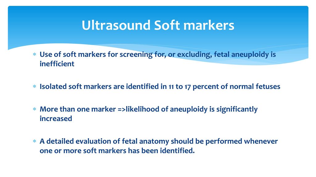
Soft markers for down syndrome on ultrasound. Most of the time this represents nothing but in some cases it may be caused by Down syndrome cytomegalovirus. Soft markersDown syndrome. Both major structural abnormalities and minor soft markers can be detected by ultrasound in fetuses affected with aneuploidies.
It also screens for neural tube defects but ultrasound has replaced this screen as a superior modality. Down Syndrome can include cardiovascular central nervous craniofacial musculoskeletal gastrointestinal and urinary tract system anomalies. Age and LR is the likelihood ratio 4.
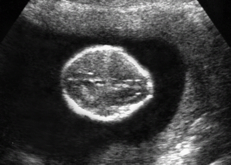
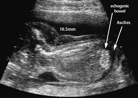
Choroid plexus cysts on the brain and an echogenic bowel. This soft marker has a higher correlation to Down syndrome. But since she saw two paired with my crappy bloodwork she was concerned about Trisomy 18.
One soft marker that might have shown up on the first-trimester NT screening which is always performed between weeks 10 and 13 is nuchal-fold thickening where the area at the back of a babys neck accumulates fluid causing it to appear thicker than usual. Thickened nuchal fold nuchal translucency Duodenal Atresia double bubble Echogenic bowel Cardiac heart anomalies Choroid plexus cyst Echogenic intracardiac focus. It is seen in approximately 20 of all Down syndrome fetuses usually in association with other findings on ultrasound.
Sometimes there can be very bright spots seen within the babys abdomen or liver. The specialist wouldnt have been as concerned if she had only seen one soft marker. Apparently these can be indicators that your baby has down syndrome.
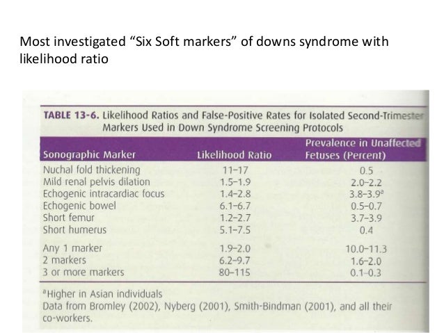
Patient-specific risk for Down syndrome O MALR 1 1LR. In the presence of soft markers the risk of Down syndrome is recalculated as new risk baseline risk x likelihood ratio LR. The ultrasound findings must be independent from one another to justify multiplying them together.
It turns out that the doctor found 2 soft markers on our ultrasound. These anomalies are not always detected by prenatal ultrasound.
One was a cyst in the babies brain.
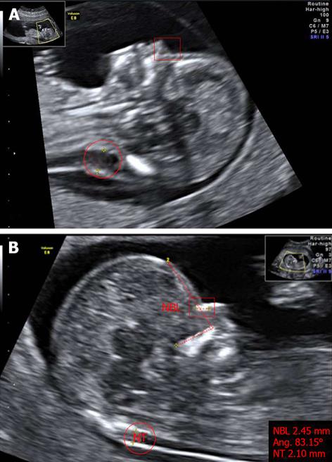
Hi guys Last week I had my 20 weeks ultrasound at 22 weeks. The following are ultrasound markers that are seen more frequently in fetuses with Down syndrome. These so-called soft markers considered as risk factors for Down syndrome and other aneuploidies were systematically described in what was called the genetic sonogram by researchers such as B. We opted for the amnio. Thickened nuchal fold echogenic bowel shortened femur shortened humerus pyelectasis and absent or hypoplastic nasal bone. The specialist wouldnt have been as concerned if she had only seen one soft marker. Importantly the test does _not_ tell you if the baby had downs it only. This soft marker has a higher correlation to Down syndrome. Certain features detected during a second trimester ultrasound exam are potential markers for Downs syndrome and they include dilated brain ventricles absent or.
But there is nothing to worry about and my child is perfectly normal Becauese my first-trimester scan and blood test were both normalBUT I need to make an appointment with a genetic. Absent or shortened nasal bone this marker has a stronger link with Down Syndrome than most others. Both major structural abnormalities and minor soft markers can be detected by ultrasound in fetuses affected with aneuploidies. But there is nothing to worry about and my child is perfectly normal Becauese my first-trimester scan and blood test were both normalBUT I need to make an appointment with a genetic. This is a relatively common finding on ultrasound. It is seen in approximately 20 of all Down syndrome fetuses usually in association with other findings on ultrasound. Choroid plexus cysts on the brain and an echogenic bowel.













Posting Komentar untuk "Soft Markers For Down Syndrome On Ultrasound"